Understanding Keratoconus
What is it, what causes it, how is it diagnosed and how is it treated and managed?
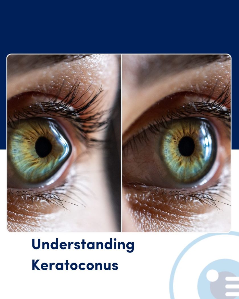
What is Keratoconus?
Keratoconus is a progressive eye disease that affects the structure and shape of the cornea, which is the clear, dome-shaped front surface of the eye.
Normally, the cornea is smooth and evenly curved, which allows it to focus light properly onto the retina for clear vision. In keratoconus, the cornea gradually thins and begins to bulge outward into a cone-like shape. This abnormal curvature causes light to scatter as it enters the eye, leading to distorted and blurred vision.
Keratoconus often begins during the teenage years or early twenties and may progress for 10 to 20 years before stabilising. It typically affects both eyes, although the degree of progression may vary between them.
Causes of Keratoconus
The exact cause of keratoconus remains unclear, but medical research has concluded it is due to a combination of genetic, environmental, and biochemical factors as detailed below.
- Genetics: Around 10-15% of people with keratoconus have a family history of the condition. Specific genetic mutations may make the cornea more susceptible to weakening and thinning over time.
- Enzymatic imbalance: Some studies suggest that people with keratoconus have an imbalance of enzymes in the cornea. This imbalance may reduce protective antioxidants in the corneal tissue, leading to oxidative damage and structural weakening.
- Eye rubbing: Chronic or forceful eye rubbing is a significant risk factor for keratoconus. It may contribute to the mechanical breakdown of the corneal tissue, especially in individuals already predisposed to the condition.
- Allergies or allergic immune responses: People with conditions like asthma, eczema, or hay fever may be more likely to develop keratoconus. These conditions often cause itchy eyes, leading to increased eye rubbing.
- Underlying medical conditions: Keratoconus has been associated with systemic diseases such as Down Syndrome, Ehlers-Danlos Syndrome (EDS) and others.
Detection and Diagnosis
Early detection of keratoconus is critical for managing the disease and preventing serious visual impairment. Diagnosis usually involves a comprehensive eye examination, which may include the following tests:
- Visual acuity test: Measures how well a person can see letters or symbols from a distance. People with keratoconus often experience worsening visual acuity that cannot be fully corrected with glasses, presenting an early indicator.
- Corneal topography: The most accurate and commonly used test to detect keratoconus. This computer-assisted procedure creates a detailed map of the curvature of the cornea and can detect even early changes in corneal shape.
- Corneal pachymetry: This measures the thickness of the cornea. Thinning of the cornea, especially at the apex of the cone, is a hallmark of keratoconus and presents an indicator of the disease.
- Slit-Lamp examination: a microscope with a bright light, which examines the front part of the eye. In moderate to advanced keratoconus signs like Fleischer rings (iron deposits) or Vogt’s striae (fine stress lines) may be visible.
- Keratometry: Measures the curvature of the anterior corneal surface using reflected light. While not as precise as topography, it can help monitor changes in corneal shape over time.
Management and Treatment Options
Keratoconus cannot be cured, but a range of management options exist to slow its progression and improve vision. The choice of treatment depends on the stage and severity of the disease and will be advised by your ophthalmologist.
- Glasses and soft contact lenses: In the early stages of keratoconus, vision can often be corrected with prescription glasses or soft contact lenses. These methods help compensate for mild irregular astigmatism. However, as the disease progresses and corneal distortion worsens, glasses and soft lenses may no longer provide adequate vision correction.
- Rigid gas permeable (RGP) contact lenses: These hard lenses maintain a fixed shape and rest on the irregular cornea, creating a smooth refractive surface. RGP lenses are effective for moderate keratoconus but require careful fitting and patient adaptation.
- Scleral and Hybrid Lenses: Scleral lenses are large-diameter gas-permeable lenses that vault over the cornea and rest on the sclera (white part of the eye). They are highly effective for advanced keratoconus because they provide comfort and excellent vision even over a distorted cornea. Hybrid lenses combine a gas-permeable center with a soft outer skirt to improve comfort and stability.
- Corneal Cross-Linking (CXL): This is the only treatment proven to halt the progression of keratoconus. It involves applying riboflavin (vitamin B2) eye drops to the cornea and then exposing it to ultraviolet-A (UV-A) light. This process strengthens the chemical bonds in the corneal collagen, making the cornea more stable and resistant to further bulging. CXL is usually recommended when there is evidence of progression, particularly in younger patients. It does not improve vision directly but can be combined with other therapies for better outcomes.
- Intacs (Corneal Inserts): These are small, curved implants inserted into the cornea to flatten its shape and improve vision. Intacs may be helpful in mild to moderate keratoconus when contact lenses are not tolerated. They can also delay or reduce the need for corneal transplantation.
- Topography-Guided Photorefractive Keratectomy (PRK): In selected cases, especially when the disease is stable after cross-linking, topography-guided PRK can be used to improve corneal shape and vision. It involves reshaping the cornea with a laser to reduce irregularities, although it is not suitable for all patients.
- Corneal Transplantation: When keratoconus becomes severe and other treatments fail to provide functional vision, a corneal transplant may be necessary. There are two main types:
-
- Penetrating keratoplasty (PK): A full-thickness corneal transplant where the entire central cornea is replaced.
- Deep anterior lamellar keratoplasty (DALK): Replaces only the front and middle layers of the cornea, preserving the patient’s healthy endothelium. DALK has a lower risk of rejection and better long-term outcomes than PK in suitable cases.
- Success rates for this surgery and good and recovery from corneal transplant surgery can progress over months, and visual outcomes can vary.
Ongoing care for those with Keratoconus
With early diagnosis and modern treatments like CXL and scleral lenses, most people with keratoconus can maintain functional vision and avoid the need for corneal transplant.
Regular monitoring by an eye care professional remains important, especially during the disease’s most active years.
Lifestyle Considerations and how to keep your eyes healthy
People with keratoconus should avoid rubbing their eyes and manage any underlying allergies that may be relevant.
Wearing protective eyewear when relevant may be advised to prevent potential eye trauma.
As the condition can be hereditary, family members may consider screening, especially during adolescence to ensure early diagnosis and management is possible.
Looking to the future
Keratoconus is a complex but manageable eye disease that can impact clear vision by changing the cornea’s shape. While its precise cause is often due to multiple factors, a combination of genetic and environmental factors are likely to play a role. Early detection through corneal imaging and timely intervention with cross-linking and specialty lenses can greatly improve visual outcomes for those affected. Advances in treatment have transformed keratoconus from a potentially sight-threatening disease into a condition that, with proper care, can be effectively controlled.
Would you like to be screened for Keratoconus?
Dr Simone Beheregaray is a consulting ophthalmologist at Adelaide Eye & Laser Centre and has expertise and skill in the management and treatment of corneal and ocular surface diseases including in keratoconus, corneal cross-linking, and all forms of corneal transplantation.
If you would like a referral to her care or have any questions, please do not hesitate to speak with your optometrist or contact a member of our friendly team directly on 08 8274 7000 or by filling out the contact form here.